ALS and FTD are each treated as one disease. The biology suggests each is several.
Patients who look identical at the bedside often have very different cellular biology underneath. That is the leading explanation for why drug trials targeting single mechanisms have repeatedly failed. A treatment matched to one form of ALS cannot help a patient whose disease runs on a different one.
This project is building a research platform that can sort patients by their biology during life. Until now, most ALS subtype information has come from postmortem tissue. FTD exists on a spectrum with ALS, and the same biological subtypes likely cut across both diseases.
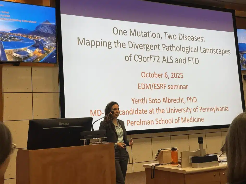
We hypothesize at least four biological subtypes:
- Iron-dominant: regional iron (or alternative metal) buildup in motor regions of the brain and spinal cord
- TDP-43-dominant: misfolding of TDP-43, the protein that normally helps cells handle their RNA and is found mislocalized in roughly 97% of ALS cases and about half of FTD cases.
- Neuroinflammatory: the brain’s own immune cells turning against motor neurons
- Oxidative lipid stress: damage to the fats that make up cell membranes, often layered on top of one of the above
To find these subtypes, the platform combines four kinds of measurement. A low-field MRI scan in development detects iron in motor regions of the brain and spinal cord. Synchrotron beamlines at the ESRF in Grenoble add the ability to map other metals in donated brain and spinal cord tissue, as well as patient cells. A blood-based assay (in development) measures the harmful, mislocalized form of TDP-43. A second blood assay measures damaged cell-membrane fats. AI analysis of patient samples sifts through thousands of proteins and lipids to find recurring patterns that distinguish one subtype from another. Each measurement on its own gives a partial picture; combined, they begin to give a fingerprint.
The same platform also aims to produce pharmacodynamic biomarkers. These are tests that show whether a candidate drug is hitting its biological target in a patient. The information arrives early enough to course-correct a trial. Pharmacodynamic biomarkers are what make precision-medicine trials possible, including preclinical antibody therapies that aim to clear mislocalized TDP-43 and approaches targeting oxidative lipid damage. None of these tools are clinically validated yet. They are research tools under active development.
Why this matters for C9 carriers
C9orf72 is the most common genetic cause of ALS and FTD, and the same mutation produces ALS in some carriers, FTD in others, both in some, and (occasionally) neither. A platform that can resolve which biological pathway is driving disease in a living patient gives the C9 community a way to sort carriers by likely trajectory, time interventions when they make biological sense, and test C9-specific therapies in the patients most likely to respond.
If I am not able to cure myself in time, this is the platform that, which time, could tell my doctors which form of ALS or FTD I am developing — and which therapy has the best chance of helping.
Principal investigators
Prof. Jenna Gregory, Neuropathologist (MD, PhD), University of Aberdeen.
- Lab: https://gregorylaboratory.com/
- LinkedIn: linkedin.com/in/jenna-gregory7
Dr. Pegah Masrori, Neurologist (MD, PhD), Institute of Neuroscience, UCLouvain.
- LinkedIn: linkedin.com/in/pegahmasrori
Partners
- European Synchrotron Radiation Facility (ESRF)
- Scientists at UCLouvain
- Scientists at University of Aberdeen
- Scientists at University of Porto
- Scientists at University of Bordeaux
- Scientists at Utrecht University
- Target ALS (sample and antibody provision)
What this project does
- Develops low-field MRI sequences sensitive to iron buildup in motor regions of the brain and spinal cord
- Maps which metals accumulate on the abnormal protein and RNA aggregates caused by C9 expansion, and how that changes as disease progresses.
- Develops blood-based assays for mislocalized TDP-43 and damaged cell-membrane fats
- Uses AI to integrate imaging, protein, lipid, and immune data into candidate fingerprints for distinct ALS subtypes
- Tests subtype-specific biology and candidate therapies in patient-derived organoids — small three-dimensional tissues grown from patients’ own cells
- Builds biomarkers usable for long-term tracking and for precision-medicine trials, where the right drug is given to the right patient based on biology, not bedside symptoms
What this project needs
- Funding.
- Well-characterized post-mortem brain and spinal cord tissue from genetically defined ALS and FTD cases, especially C9orf72 carriers.
- Larger-scale validation. The imaging and AI pipelines are built. Running them across hundreds of patients is what turns four candidate subtypes into a tool the field can rely on.
